
Lub dub…lub dub…lub dub…
Can you hear that? It’s the beating of a heart. You can’t deny that there is something enchanting about the rhythmic beating of a human heart. This organ, responsible for pumping blood throughout the body 24/7, is by far one of the most vital within the body. As you can probably assume by now, its time to study the heart in my Anatomy and Physiology class! Whooo!
To get the blood flowing (pun definitely intended!) with this new unit, we started with a heart dissection.However, this wasn’t like any normal dissection. You see, the class was distributed sheep and pig hearts to cut open. These by themselves aren’t very large in size. My group, however, was an exception. We were given something bigger.Something way more interesting- we were given a cow heart! Let me just put the size of a cow’s heart in perspective. Below are my good colleagues Trevor Lopkoff and Chuck Aaron, posing with the organ:

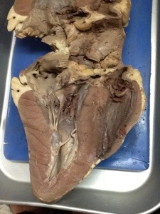
Holy cow! (Yes, I intended that one also!). See what I mean when I say it’s huge? It fills the entire dissection tray! For this dissection, we cut the heart along the frontal plane, meaning we cut it down the middle and separated the anterior side from the posterior side. This way, we are able to see the chambers, valves, and inner components of the heart much more clearly! Take a look!

The purpose of this lab was to learn theanatomical structures of the heart, while also comparing the sizes of the hearts of different animals. We ended up taking some measurements on the cow heart. In short, they are listed below:
Aorta: 2.5 cm thick
Left Atrium: 10 cm thick
Left Ventricle: 2.5 cm thick
Right Atrium: 8 cm thick
Right Ventricle: 3.5 cm thick
Outer Wall: 2.5-3 cm thick
Now, for comparison purposes, let’s look at the measurements of a sheep’s heart, as provided by another group in my class:
Aorta: 1 cm thick
Left Atrium: 3.5 cm thick
Left Ventricle: 2 cm thick
Right Atrium: 4 cm thick
Right Ventricle: 0.75 cm thick
Outer Wall: 1.5-2 cm thick
You don’t have to be a mathematician to realize just how significant the discrepancy is between these two animals! It is truly amazingto think that a heart, nearly identical in appearance to a human, can be so much larger than ours! That’s incredible!
After the excitement of dissection was over, it was time to learn about the components of the heart. For this post, I will provide only the basics. After all, the heart is such a complex organ! To explain the components of the heart, I will be using a diagram of a heart that was labeled by yours truly! (It was technically a quiz for my class, but I aced it!). A secondary diagram is included below the description in case my quiz is too difficult to read.

The heart is divided into four main chambers: the Right Atrium, Right Ventricle, Left Ventricle, and Left Atrium. The heart also has four valves: the Tricuspid Valve (right side), the Pulmonary Valve (right side), the Aortic Valve (left side), and the Mitral Valve (left side).
The heart works in the following way: De-oxygenated blood that has been circulating through the body gets returne
d through the Vena Cava (the Superior Vena Cava receives blood from the upper part of the body, while the Inferior Vena Cava receives the blood from the lower half). This “used” blood enters the Right Atrium. The Atrioventricular (AV) and Sinoatrial (SA) Nodes are located here, and are designed to help maintain a rhythm for the heart to pump blood with.
From here, the blood passes through the Tricuspid Valve and enters the Right Ventricle. The next step is to pass through the Pulmonary Valve, and enter the Pulmonary Artery. This pathway leads directly to the lungs, where blood passes through arterioles, capillaries, and venules (in that order) to obtain oxygen. After getting oxygen from the lungs, the blood is reinserted into the heart through the Pulmonary Vein.
The Pulmonary Vein releases blood into the Left Atrium. Then the blood passes through the Mitral Valve and enters the Left Ventricle. The final step is to pass through the Aortic Valve and enter the Aorta, which distributes the blood through the rest of the body.
Also, it is important to note that the muscle that separates the two sides of the heart (between the right and left ventricules) is called the Septum.

With that, we have now achieved the two main goals for this post: we have learned about the heart through a detailed explanation
of how it works, and we have seen it on a grand scale through an awesome dissection. The most important thing to realize is that the heart is the muscle that works constantly to keep the blood in the body circulating. It is a vital part of the human body! And to study it, might I add, is something you just can’t beat! (Get it?)
